Archives

Retinal Vein Occlusions

Retinal Vein Occlusions
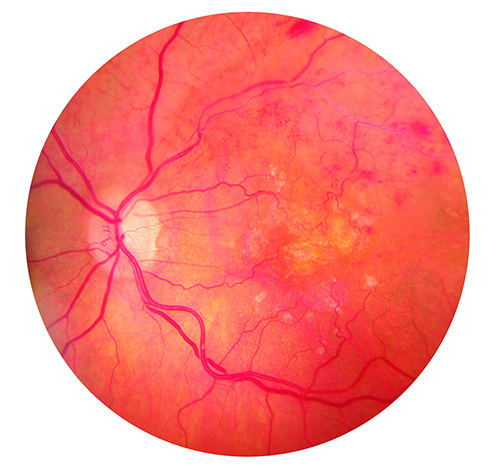
The retina is the nerve tissue in the back of the eye that requires healthy blood circulation for visual function. When blood vessels to the retina are blocked, it is known as a retinal vein occlusion or “eye stroke” and can cause sudden blindness.

There are two types of retinal vein occlusions: Central Retinal Vein Occlusion (CRVO) and Branch Retinal Vein Occlusion (BRVO).

About

CRVO

Blood enters the retina through the central retinal artery and drains out of the retina through the central retinal vein. When the central retinal vein becomes blocked, this is called a central retinal vein occlusion or CRVO. This means that blood cannot flow in and out of the retina properly, causing the retina to fill up with blood and swell, resulting in vision loss.

There are two types of CRVO:

  • Non-ischemic CRVO. This is less serious and accounts for a majority of the cases.
  • Ischemic CRVO. This is the more serious form because the retina becomes so “starved” for oxygen that new blood vessels grow in various locations in the eye to improve the insufficient blood supply. Unfortunately, this is dangerous and can lead to vision loss and possibly loss of the eye.

BRVO

This is when the small branches of the main vein in the retina are blocked. As with CRVO, part of the retinal blood flow slows down or stops which results in vision loss. This usually occurs in one eye and results in blurred vision or a missing area of vision.

Symptoms

  • Sudden loss of vision or blurring in part or all of one eye
  • Floaters (small moving spots) in your vision in severe cases
  • Painful pressure in the eye

Prevention

As in other health issues a healthy diet and lifestyle help stave off Retinal Vein Occlusion. In addition, to that maintaining diabetes is also helpful.

Diagnosis

Diagnosing eye conditions requires eye exams, which include the specialist looking for fluid or blood in the back of the eye after performing dilation and also testing for defects in central vision.

Questions to Ask Your Health Care Provider

  • Is there any chance that my vision will improve?
  • Are there treatments are available for my condition?
  • What type of tests will my eye specialist perform?
  • Will this disease spread to my other eye?
  • Will I lose my vision?
  • How will my lifestyle change after diagnosis?
  • What lifestyle changes do I need to make to delay progression of the disease?

Treatments

Unfortunately there is no way to unblock retinal veins. However it may be possible to prevent another blockage from forming in the same or the other eye. This can be done through laser treatment, drug injections or a vitrectomy (surgery). Many people may regain some of the lost vision without treatment, however, their vision rarely returns to normal.

Last modified: June 4, 2014

Glaucoma

Diabetic RetinopathyGlaucoma is a group of eye disorders that can damage the eye’s optic nerve and eventually result in vision loss or blindness. It develops slowly and sometimes without noticeable sight loss for many years. However, with early detection and treatment, it is possible to protect your eyes from a serious loss of vision.

About

Risk factors for glaucoma include:

  • being over 40
  • being African-American or Hispanic
  • family history of glaucoma
  • being diabetic

Because most people with glaucoma have no early symptoms or pain from this increased pressure, it is important to see your eye doctor regularly so that glaucoma can be diagnosed and treated before long-term visual loss occurs.

Glaucoma occurs when pressure in your eye increases due to a lack of circulation of the eye fluid in the front part of the eye.

FYI

Glaucoma is the second most common cause of blindness in the U.S. There major types of glaucoma are:

  • Open-angle glaucoma (most common type, and happens slowly over time)
  • Closed-Angle glaucoma (sudden fluid blockage and a quick rise in pressure)
  • Congenital glaucoma (occurs in babies)

Symptoms

  • Loss of side vision which, in advanced stages, can develop into tunnel vision
  • Sudden, severe blurring of vision
  • Severe pain in or around the eye
  • Eye redness
  • “Halos” around lights

Prevention

You can’t prevent glaucoma and most people have no symptoms, but you can help prevent vision loss.

  • All adults should have a complete eye exam by the age of 40.
  • If you are at risk for glaucoma, you should have a complete eye exam sooner than age 40.
  • You should have regular eye exams as recommended by your doctor.
  • If you are at risk for closed-angle glaucoma, your doctor may recommend treatment before you have an attack to help prevent eye damage and vision loss.

Diagnosis

Diagnosing eye conditions requires eye exams, which include the specialist looking for fluid or blood in the back of the eye after performing dilation and also testing for defects in central vision. The specialist will also perform a pressure test where a machine blows a puff of air into the eye.

Questions to Ask Your Health Care Provider

  • What type of glaucoma do I have?
  • How will my vision be affected now and in the future?
  • Is it hereditary? What should I tell my family about my condition?
  • What is my expected prognosis?
  • Is it safe for me to drive?
  • Is this hereditary?
  • Will I lose my vision?
  • What are my treatment options?
  • How often do I need to have check-ups once I am diagnosed?

Treatments

Early diagnosis is important as it can delay progression of the disease. Although treatment may save your remaining vision, it does not improve sight already lost from glaucoma.

Treatments include

  • Medicines: This is usually in the form of eye drops or pills. These lower your eye pressure by making the eye less fluid or draining fluid from the eye.
  • Laser trabeculoplasty: This surgical procedure helps to drain fluid out of the eye.
  • Conventional Surgery: This makes a new opening for the fluid to leave the eye. This is often done if other methods do not work.

Your healthcare provider may also use a combination of the treatments mentioned above.

Last modified: June 2, 2014

Cataracts

CataractsA cataract is when the clear lens of your eye is cloudy. Having cataracts can create difficulty reading, driving a car — especially at night or seeing the expression on a person’s face.

About

What causes cataracts?

The lens lies behind your eye’s iris and pupil. This lens focuses light onto the retina at the back of your eye where an image is displayed. The lens also functions to adjust the eye’s focus, which allows you to clearly see things near point and at a distance. Lenses consist of mostly water and protein. That protein is precisely arranged in a way to keep the lens clear — letting light pass through it.

During the aging process, some of the protein can clump together creating a cloud (cataract) on a small area of the lens. Eventually the cataract may increase in size and cloud more of the lens, which makes vision problematic.

It is suspected in the scientific field that smoking and diabetes, along with several other potential causes, make us vulnerable to cataracts. However it may also be a result of the toll the lens takes from wear and tear over the years.

Symptoms

  • Clouded, blurred or dim vision
  • Increasing difficulty with vision at night
  • Sensitivity to light and glare
  • Seeing “halos” around lights
  • Frequent changes in eyeglass or contact lens prescription
  • Fading or yellowing of colors
  • Double vision in a single eye

Prevention

Maintaining regular exams with your eye care specialist will ensure that you’re monitoring your vision properly. The optometrist will be able to note changes in vision between each appointment. You must also control diseases that increase the risk of a cataract and avoid exposure to things that promote cataract formation. It’s always beneficial to wear sunglasses to protect your eyes from harmful UV rays when outdoors.

Diagnosis

Eye exams are done with a slit-lamp exam. An optometrist will shine a light into your eye and looking through a scope, be able to see in the back of your eye.

Questions to Ask Your Health Care Provider

  • How often should I see my eye doctor for routine exams?
  • My vision is sometimes blurry; does that mean I have cataracts?
  • I have a family history of cataracts, will I get them?
  • Will I experience further vision loss?
  • Will taking a vitamin or mineral supplement help prevent further vision loss?
  • What’s the best way to monitor my vision for any changes?
  • What low vision aids or adaptive devices might be helpful to me?
  • Is it safe for me to drive?
  • Can I remove cataracts?

Treatments

During the early stages, the effects of cataracts may be improved with new eyeglasses, brighter lighting, anti-glare sunglasses or magnifying lenses. Surgery is the only successful treatment if none of the previously mentioned measures help. This involves the removal of the cloudy lens and replacing it with an artificial lens. Cataract patients may also have other age-related eye conditions, such as macular degeneration or glaucoma. Be sure to talk to your healthcare provider about how these conditions may be related.

Last modified: June 2, 2014

Age-Related Macular Degeneration (AMD)

Age Related Macular Degeneration (AMD)

This is the leading cause of vision loss in people aged 50 years and older. It happens when a small central portion of the retina, known as the macula, deteriorates. The macula is the part of the eye that provides sharp, central vision needed for seeing objects clearly. Having this condition makes it harder to recognize faces, walk, drive, read or do work close up like fixing things around the house.

About

There are two main types of AMD: Dry and Wet.

DRY AMD

This is when the light-sensitive cells in the macula slowly break down, gradually blurring central vision in the affected eye. This is the more common of the two types of AMD. In the early stages of Dry AMD, you may not be aware of any symptoms. As the disease advances, you may notice the following:

  • Visual distortions of objects, such as straight lines appearing crooked or wavy, or objects such as street signs appearing lopsided
  •  Objects appearing smaller than they are, or father away
  • Decreased vision in the center of your viewing area
  • Colors appearing dull or lessened in intensity
  • Blurry or blind spots in your field of vision
  • Seeing shapes, people and /or animals that are not really there

WET AMD

Wet AMD is the more advanced type of AMD. This is when abnormal blood vessels develop beneath the macular retina and leak blood and fluid, resulting in scarring and visual impairment. Wet AMD usually progresses more rapidly than the dry version, and generally leads to more serious vision loss.

FYI

Macular degeneration is most common in people older than 65.

RISK FACTORS

  • Age. Risk increases as you age, especially after age 50
  • Family history of macular degeneration
  • Race. Macular degeneration is more common in Caucasians than it is in other races
  • Smoking
  • Obesity
  • Diet. A diet that includes few fruits and vegetables may increase the risk of macular degeneration
  • High blood pressure. Diseases that affect the circulatory system, such as high blood pressure or high cholesterol, may increase the risk of macular degeneration
  • Inflammation. Your immune system can cause swelling of your body tissues, which may increase the risk of macular degeneration
  • Cardiovascular disease

Symptoms

DRY AMD Symptoms

  • Needing brighter light when reading or doing close work
  • Difficulty adapting to low light levels, such as when entering a dimly lit room
  • Increasing blurriness of printed words
  • A decrease in the intensity or brightness of colors
  • Difficulty recognizing faces
  • Hazy or less well-defined vision
  • Straight lines look irregular or bent, and objects appear a different color or shape in each of the eyes

WET AMD Symptoms

  • Visual distortions, such as straight lines appearing wavy or crooked, a doorway or street sign looking lopsided, or objects appearing smaller or farther away than they really are
  • Decreased central vision
  • Decreased intensity or brightness of colors
  • Blurry spots or blind spots in your field of vision
  • Seeing shapes, people and /or animals that are not really there

Prevention

  • Routine eye exams
  • Manage your other diseases
  • Stop smoking
  • Maintain a healthy weight and exercise regularly
  • Choose a diet rich in fruits and vegetables, and include fish

Diagnosis

Eye exams are done with a slit-lamp exam. An optometrist will shine a light into your eye and looking through a scope, be able to see in the back of your eye.

Questions to Ask Your Health Care Provider

  • How often should I see my eye doctor for routine exams?
  • What kind of macular degeneration do I have?
  • How advanced is my macular degeneration?
  • Will I experience further vision loss?
  • Will taking a vitamin or mineral supplement help prevent further vision loss?
  • What’s the best way to monitor my vision for any changes?
  • What low vision aids or adaptive devices might be helpful to me?
  • Is it safe for me to drive?

Treatments

Macular degeneration can’t be cured and treatment can’t reverse what’s already occurred. Dry macular degeneration usually progresses slowly and many people with the condition can live relatively normal, productive lives especially if only one eye is affected. In either case, if diagnosed early, you can do things to help slow progression.

DRY AMD Treatment

There is currently no cure for Dry AMD, but there are steps you can take that may reduce vision loss. This includes changes in your diet and vitamin intake. Your healthcare provider may also recommend surgery to implant a telescopic lens in your eye to improve your vision.

WET AMD Treatment

Wet AMD can result in severe vision loss. However, treatments from your eye care professional may prevent further vision loss. These treatments can include injections of drugs that block the growth of abnormal blood vessels. Less commonly, laser surgery may be performed to treat Wet AMD.

Last modified: June 2, 2014

Diabetic Retinopathy

Retinal Vein OcclusionsThis is the most common diabetic eye disease. When diabetes is untreated, it can damage the small blood vessels in your retina, which is located in the back part of your eye. The damaged blood vessels cause bleeding, swelling and scarring that can lead to poor vision or blindness. This is called diabetic retinopathy. You usually only notice symptoms once the damage is severe.

About

Anyone who has Type 1 or Type 2 diabetes has the potential to develop diabetic retinopathy. The longer you have diabetes, and the less controlled your blood sugar is, the more likely you are to develop this serious vision problem.

Diabetic retinopathy can result in blindness.

Symptoms

  • Blurred vision and slow vision loss over time
  • Shadows or missing areas of vision
  • Trouble seeing at night
  • Difficulty with color perception

Prevention

To prevent diabetic retinopathy you must begin by controlling your diabetes, blood sugar levels and making annual eye exams. You also need to pay attention to your vision between appointments, making sure you note any sudden or severe changes.

Diagnosis

As in any eye exam, the healthcare provider will dilate your eyes for a clear view into the eye. The provider will look for abnormal swelling, blood or dark fluid. They will also monitor your glucose levels from your primary healthcare provider’s notes on file. You may be required to have blood work done prior to the appointment.

Questions to Ask Your Health Care Provider

  • Why is diabetes affecting my eye sight?
  • What could I do in addition to maintaining my blood sugar levels to stop this disease?
  • How often should I see my eye doctor for routine exams?
  • Will I experience further vision loss?
  • Will taking a vitamin or mineral supplement help prevent further vision loss?
  • What’s the best way to monitor my vision for any changes?
  • What low vision aids or adaptive devices might be helpful to me?
  • Is it safe for me to drive?
  • What types of side effects could I have from the treatments?
  • If I control my diabetes, will this problem go away?

Treatments

To prevent the disease from getting worse during the early stages, you should control your levels of blood sugar, blood pressure, and cholesterol .

During the later stages, although there is no cure, your healthcare provider may recommend getting scatter laser treatment or a vitrectomy (surgery) to reduce vision loss.

Last modified: June 2, 2014